The document was formerly published as GEX document number 100-211.
Purpose
The intent of this document is to provide low energy electron beam dosimetry users with a status update on the new Dµ method of calibration introduced in November 2007 by GEX and Risø High Dose Reference Laboratory that utilizes reference transfer standard alanine film dosimeters to provide doses traceable to a national standard.
Dµ Calibration Process
The Dµ calibration process was designed to give low energy electron beam users a method for the traceable calibration of routine dosimetry systems and conforms with existing GEX published practices for “in-situ” or “in-process” calibration of routine dosimetry systems as well as ISO/ASTM standards for dosimetry system calibration. For the purposes of this document, we define low energy to be electron energies of 300 keV or less
Risø National Laboratory supplies GEX with the transfer dosimeters (130 micron nominal thickness alanine films) which are sent to customers who irradiate them alongside their routine B3 radiochromic dosimeters using protocols specified by Risø and GEX. The customer returns the alanine film dosimeters to Risø who measure the alanine films and correct the measured doses for the specific in-plant conditions of the user’s machine and facility conditions.
The transfer alanine dosimeter results are corrected to account for the actual beam energy penetration at the dosimeter surface as actually measured in a depth/dose stack. Other correction factors are used by the laboratory to account for the user’s stated distance between the external surface of the accelerator window and dosimeter surface (air gap) as well as the accelerator window material composition and its thickness. Additionally, the laboratory makes a temperature correction based on an estimated average temperature in the alanine dosimeters during irradiation.
The calculated surface dose in the transfer alanine dosimeters is designated as a Dµ dose and is considered to be the average dose in the first micron of the transfer alanine dosimeter.
GEX combines the reported customer’s B3 batch calibration dosimeter measurements with the Risø reported Dµ doses to develop the calibration response function for the routine dosimeters in terms of Dµ dose that is traceable to a national standard with a stated level of uncertainty.
Value of Dµ Calibration Method
The new Dµ calibration method resolves the historical inability to establish calibration traceability to a national standard through an unbroken chain of calibration events for dosimetry systems used in low energy electron beam applications. For the first time, this new method provides the low energy user with traceable transfer standard doses equivalent to those that have been available and widely used by gamma and high energy e-beam users for more than twenty years.
Low Energy Dosimetry Calibration Background
For many years, low energy users relied on the use of a gamma source calibration performed at laboratories using known dose rate gamma irradiations. Unfortunately, these were fixed temperature irradiations that did not take into account the impact of temperature influence on the response of the routine dosimeters that occurs when they are used in production facilities where the high dose rates of the electron beam causes a significant rise in temperature to the dosimeter during irradiation. This technical issue coupled with unknown environmental events during shipments to and from remote locations along with post irradiation response measurement changes led to inaccurate dosimeter measurements by the end user.
GEX recognized the need to eliminate the bias associated with the use of a laboratory gamma calibration in low energy processing many years ago and developed a practice of performing high energy electron beam calibrations through Risø. These high energy electron calibrations provide more accurate calibrations than the original gamma method because they accounted for the influence of temperature on the response function of the dosimeter by capturing temperature influence in the calibration irradiations.
However, calibration of dosimetry systems using the high energy electron beam method can introduce batch to batch differences associated with change to the calibration practice (e.g. temperature).
Another challenge was to determine the true response function differences between a calibration function derived from a high energy radiation source and the true response function of the film in a low energy source. The question was partially answered with investigations carried out and published in 2005 by Miller, Helt-Hansen, Sharpe, et al using specially developed low energy calorimeters which demonstrated agreement within 10% of comparable 10 MeV electron calibrations.
However, it was not until the Dµ method became available that the question could be addressed by using doses traceable to a national standard at a known level of uncertainty. The new Dµ doses allow the low energy user to perform audit verification of high energy electron beam calibrations or the ability to perform a fully traceable in-situ calibration and compare the differences.
At this time however, only limited field testing has been carried out in an effort to validate the new method. These results were presented formally by Risø at the September 2008 International Meeting on Radiation Processing (IMRP) and will be subsequently published. Additional activities have included peer review discussions with open presentations made at the past two meetings of the ASTM E10.01 Sub-Committee on Dosimetry as well as at the Gamma and Electron Radiation Panel workshop on Advanced Dosimetry Techniques immediately following the IMRP 2008 meeting. The initial rounds of field tests involving the new Dµ doses have provided reproducible results.
The following Technical Appendix shows the results obtained by GEX using this new Risø Dµ method in a series of low energy calibrations. Also provided is a discussion of the issues surrounding low energy electron beam dosimetry.
Summary
For the first time, a truly traceable calibration method has been developed and tested for dosimetry systems used in low energy electron beam irradiation. Batch specific dosimetry system calibration for use in low energy electron beam applications can now be carried out using an “in-situ” or “in-process” calibration method in the user’s plant as described in the ISO/ASTM 51261 document. A method has been developed and tested where routine dosimeters are irradiated together with reference transfer dosimeters to the same doses using low energy electrons. The transfer doses are stated by the laboratory in terms of a surface dose called Dµ. A calibration response function is derived in terms of routine dosimeter response versus the laboratory reported Dµ doses and this response function is used to estimate doses from measurements made using the customer’s routine dosimetry system.
New Dµ traceable doses can be used to verify use of a 10 MeV calibration for use in low energy electron beam irradiations. It can be demonstrated that a generic 10 MeV calibration may be used with an approximate ±7.0% or better agreement with Dµ doses at surface energies down to approximately 90 keV (approximately 125 kV accelerator energy set point) when using a thin B3 dosimeter that is nominally 17 microns thick. The data indicates that dosimetry calibration for dosimeter surface energies below 90 keV (~125 kV accelerator energy set point) require a full in-situ calibration using the Dµ method to obtain any degree of dose measurement accuracy.
Technical Appendix
This appendix reports on the results of initial experience using the new Dµ calibration method for the calibration of dosimetry systems used in low energy electron irradiation applications. Both Risø and GEX have established procedures and work instructions for the Dµ calibration activities.
The Dµ calibration method evolved over 7 years with a significant amount of technical research needed to resolve the complexities associated with dosimetry system calibration for use in a low energy electron irradiation environment.
The combined overall uncertainty associated with Dµ doses is nearly twice that of other transfer dosimetry systems used for in-process calibration in gamma and high energy electron beam applications. This is due to the higher variability associated with thin alanine film dosimeters and the added uncertainty components involved with the corrections applied by Risø.
Although it is not essential that a user possess a technical understanding of Dµ in order to use it effectively, the references provide discussion of technical information. Most routine dosimetry users can simply enjoy the accuracy and simplicity of Dµ without concern for how the doses are arrived at by the calibration laboratory, but only that the laboratory be properly accredited to certify doses traceable to a national standard.
Low Energy Electron Dosimetry Challenges
At low electron energies, the beam window material composition and thickness along with the air gap from the window to the product reduce the electron energies and, thus, have a direct impact on the penetration range of the electrons into the dosimeter and product.
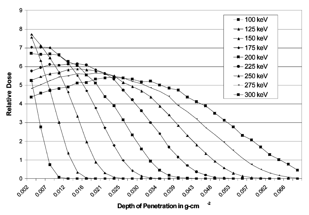
Therefore, to avoid significant variance and bias, their effects at different energy settings must be accounted for in the dosimetry system calibration process. The depth/dose plots in Figure 1 above depict the impact of energy changes below 300 keV on surface dose.
Another challenge in low energy electron beam dosimetry is the impact of depth/dose gradients occurring within the dosimeter. A ‘dose gradient’ condition in the dosimeter results when absorbed dose is not uniformly distributed throughout the thickness of the dosimeter material whereby dose builds up on the front or back surfaces of dosimeters as a result of the electron beam energy and the dosimeter thickness.
These dose gradients become more significant at lower energies (<90 keV at the dosimeter surface). At these low energies the dosimeter becomes fully absorbing of all the electron energy. Therefore, a thicker dosimeter will exhibit a steeper gradient slope than a thinner dosimeter as energy decreases.
The measured absorbance value of an irradiated dosimeter provides an “average value” representing the portion of dose actually absorbed in the dosimeter. An optimal thickness dosimeter provides absorbed dose conditions where the dose absorbed on the front surface of the dosimeter is equal to the absorbed dose on the back surface of the dosimeter. For B3 dosimeters, this typically occurs at a surface energy of approximately 165 keV (results from an approximate 200 kV accelerator voltage setting).
Surface Dose Calibration Method
A new method of calibration (Dµ) is available and provides traceability to a national standard through an unbroken chain of comparisons. The method is based on a surface dose concept called Dµ which is used by the calibration laboratory to calculate the average dose measured in the first micron of their national standard traceable transfer standard dosimeters.
The Dµ method allows the calibration laboratory to account for the impact of the accelerator window and air gap of a user’s specific irradiator system as well as to correct for their transfer alanine dosimeter thickness to arrive at average surface dose. By simply irradiating routine thin film dosimeters alongside these transfer laboratory dosimeters a user can establish a relationship in the response of their routine dosimeters to these reported Dµ laboratory doses to establish a calibration traceable to a national standard.
Comparing Dµ with 10 MeV Dose Estimates in Low Energy Electrons
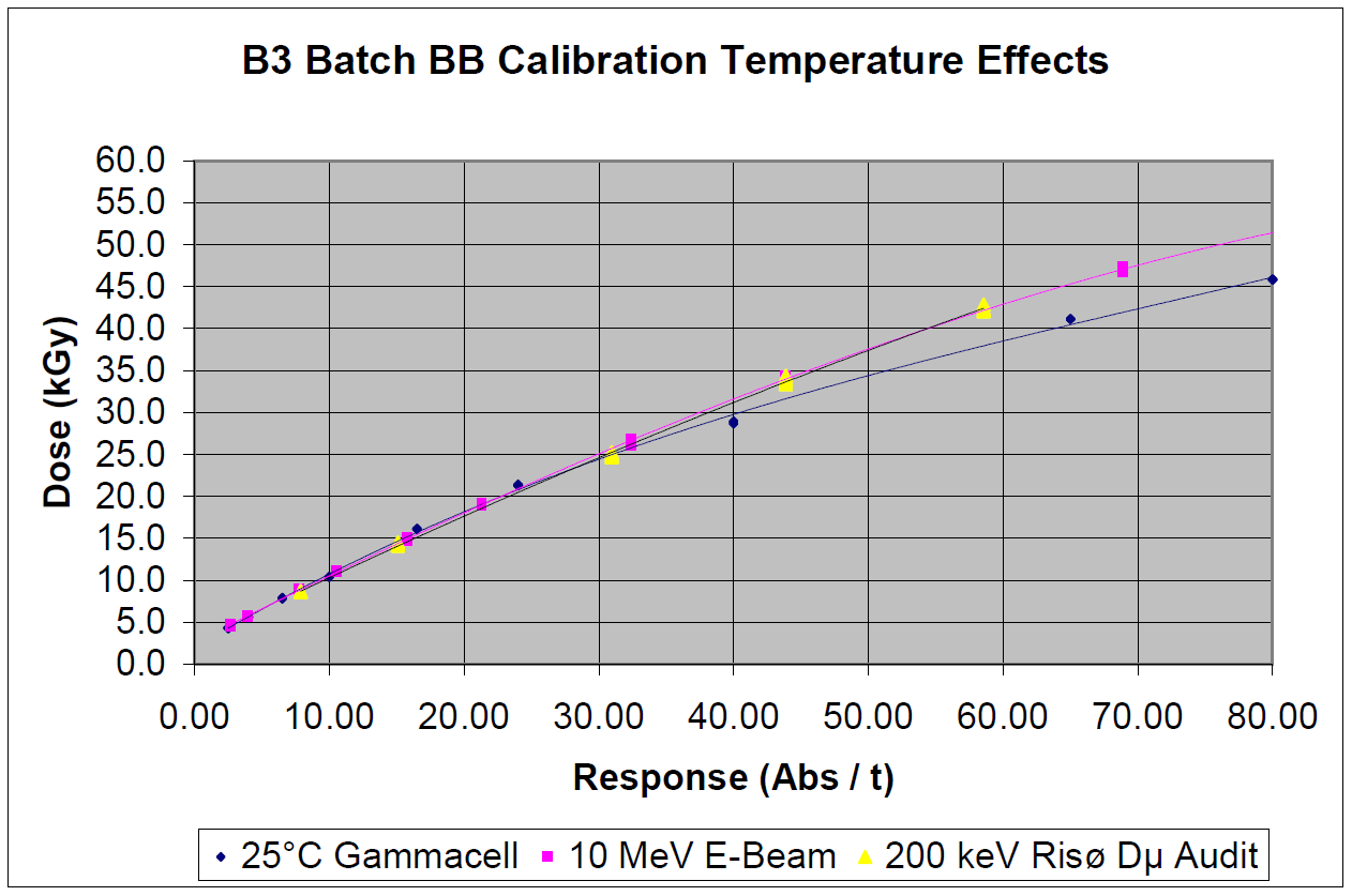
Transfer standard Dµ calibration dosimeters were used to audit batch specific laboratory calibrations of dosimeters using a high energy 10 MeV electron calibration source. The results shown in the calibration function plots below in Figure 2 demonstrate good agreement between the 10 MeV lab calibration and the national standard traceable Dµ doses.
The plots also demonstrate the clear temperature influence bias associated with the use of a gamma calibration in a low energy electron beam process. The bias would result in an overestimation of dose using a gamma calibration in a low energy irradiation process that would exceed 25% at approximately 60 kGy.
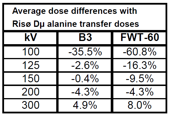
Results in the Table 1 below compare doses estimated from the Dµ calibration method using the doses estimated using a 10 MeV calibration for 17.0 micron thick B3 dosimeters and 43.5 micron thick FWT-60 dosimeters over a range of accelerator voltage settings from 100-300 kV (measured depth/dose stack energies 65-265 keV).
Conclusions
Low energy electron beam in-situ calibrations have been demonstrated to eliminate several sources of calibration bias by accounting for the impact of window, air gap, and dosimeter thickness in the calibration process while providing a means of traceability to a national standard with a similar degree of accuracy long enjoyed by gamma and high energy electron users.
References
-
Helt-Hansen, J., Miller, A., 2004. RisøScan—a new dosimetry software. Radiat. Phys. Chem. 71, 361–364.
-
Helt-Hansen, J., Miller, A., McEwen, M., Sharpe, P., Duane, S., 2004. Calibration of thin-film dosimeters irradiated with 80–120 keV electrons. Radiat. Phys. Chem. 71, 355–359.
-
Helt-Hansen, J., Miller, A., Duane, S., Sharpe, P., McEwen, M., Clausen, S., 2005. Calorimetry for dose measurement at electron accelerators in the 80–120 keV energy range. Radiat. Phys. Chem., in press, doi:10.1016/j.radphyschem. 2005.05.017.
-
Janovsky, I., Miller, A., 1987. A calorimeter for measuring energy deposition in materials and calibrating the response of dosimeters irradiated by low-energy industrial electron accelerators. Appl. Radiat. Isot. 38 (11), 931–937.
-
Kawrakow, I., Rogers, D.W.O., 2000. The EGSnrc code system: Monte Carlo simulation of electron and photon transport. Technical Report PIRS-701,NationalResearch Council of Canada, Ottawa, Canada.
-
Sharpe, P., Miller, A., 1999. Guidelines for the calibration of dosimeters for use in radiation processing. CIRM 29, National Physical Laboratory, Teddington, TW11 0LW UK.
-
Zeng, G.G., McCaffrey, J.P., 2005. The response of alanine to a 150 keV X-ray beam. Radiat. Phys. Chem. 72, 537–540.
Disclaimer -The information contained in this document is provided “as is” and is not a substitute for the user’s professional judgement. It is provided as a convenience to those using products provided by GEX Corporation who have sufficient technical skills to evaluate and properly apply the information in this document. It is the responsibility of the user of this document to ensure that the information in this document, and the use of such information, is accurate, complete, applicable to the product, suitable for the user’s purposes, and in compliance with all laws and regulations. GEX Corporation believes the information provided in this document is accurate and reliable as of the time of writing, but it undertakes no obligation to update or correct this document. GEX Corporation may, but is not required to, make changes to this document at any time without notice. By using the information in this document, the user represents and warrants that he or she has the skills necessary to properly understand and apply this information and that he or she will comply with all applicable laws and regulations including, without limitation, those relating to medical devices, pharmaceutical products, or other applicable industries. The user assumes all risks associated with using this information and any results or output resulting from the application of this information to GEX Corporation’s products. The user agrees not to hold GEX Corporation liable for any errors or omissions contained within.